Periapical X-rays play a pivotal role in the realm of dentistry, serving as an essential imaging tool that goes beyond mere diagnosis to assist in meticulous planning for dental implants. These radiographs offer a close-up view of the entire tooth—from the crown to the root nestled in the jawbone—providing invaluable insights into the bone structure and health crucial for implant success. As the cornerstone of implant planning, Periapical X-rays not only reveal the hidden details beneath the gums but also guide dental professionals in navigating the complexities of implant placement. This blog delves into the significance of Periapical X-rays, elucidating their integral function in ensuring the precision and safety of dental implant procedures.
In This Blog:
- Understanding Periapical X-Rays
- The Role of Perioapical X-Rays in Implant Planning
- Advantages of Periapical X-Rays in Implant Dentistry
- Limitations and Considerations
- Preparing for a Dental Implant Procedure
Understanding Periapical X-rays
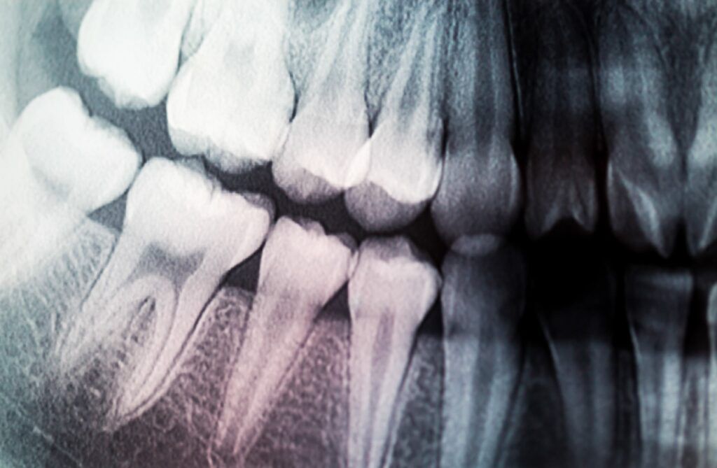
Periapical X-rays are a type of dental radiograph that provides a detailed view of the entire tooth and its surrounding bone structure. These images are critical for examining the periapex, the area around the tip of the tooth’s root, hence the name “periapical.” Unlike panoramic or bitewing X-rays, which capture a broader view of the mouth or sections of the teeth, Periapical X-rays focus on individual teeth, offering a close-up perspective that is essential for diagnosing root structure and bone health.
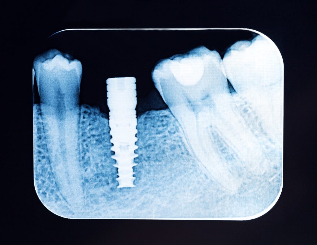
The process of taking Periapical X-rays involves placing a small film or digital sensor in the mouth, close to the tooth being examined. The X-ray machine then directs a focused beam of radiation through the tooth and onto the film or sensor, capturing a clear image of the tooth’s anatomy. This technique allows dentists to see the tooth in its entirety, from the crown to the root, along with the surrounding jawbone and soft tissues.
What sets Periapical X-rays apart from other dental imaging methods is their ability to provide a comprehensive view of the tooth and its immediate environment. While panoramic X-rays are useful for getting a general overview of the teeth and jaw, they lack the detail necessary for in-depth analysis of specific teeth. Bitewing X-rays, on the other hand, are excellent for detecting cavities between teeth but do not show the root and bone structure as Periapical X-rays do.
In summary, Periapical X-rays are a fundamental tool in dental diagnostics, offering a detailed and localized view of individual teeth and their supporting structures. This detailed imaging is crucial for assessing the tooth’s condition and the bone’s health, making it an indispensable part of the planning process for dental implants and other precise dental procedures.
The Role of Periapical X-rays in Implant Planning
Periapical X-rays serve as an essential component in the planning phase of dental implants, providing critical information that influences the entire implantation process. These detailed images are invaluable for assessing the condition of the jawbone and the health of adjacent teeth, which are key factors in determining the feasibility and approach for implant placement.
Assessing Bone Health and Structure
One of the primary uses of Periapical X-rays in implant planning is evaluating the quantity and quality of the jawbone. The success of a dental implant largely depends on the bone’s ability to support the implant post. These X-rays offer a clear view of the bone density and volume, allowing dentists to assess whether there is sufficient bone to anchor the implant securely. In cases where bone loss has occurred, the X-ray can help in planning additional procedures, such as bone grafting, to create a solid foundation for the implant.
Identifying Anatomical Landmarks and Potential Obstacles
Periapical X-rays are instrumental in mapping out the anatomical landmarks within the jaw, such as nerves, sinuses, and blood vessels. Precise knowledge of these structures is crucial to avoid complications during the implant procedure. For example, the X-ray can reveal the proximity of the inferior alveolar nerve in the lower jaw, helping to prevent nerve damage during implant placement.
Furthermore, these X-rays can detect potential obstacles such as impacted teeth, cysts, or other abnormalities that might complicate the implant process. Identifying these issues early allows for the necessary adjustments in the treatment plan, ensuring a safer and more effective implant procedure.
Periapical X-rays are fundamental in the preparatory stages of dental implant procedures. They provide detailed insights into the oral anatomy, enabling dentists to make informed decisions and plan effectively for implant placement. By thoroughly evaluating the bone structure and identifying potential challenges, these X-rays help ensure the longevity and success of dental implants.
Advantages of Periapical X-rays in Implant Dentistry
Periapical X-rays are a cornerstone in the field of implant dentistry, offering several advantages that make them indispensable in the planning and execution of dental implants. Their high-resolution images and detailed views of the tooth roots and surrounding bone structure provide a wealth of information that is crucial for successful implant placement.
High-Resolution Images for Detailed Analysis
The primary advantage of Periapical X-rays lies in their ability to produce high-resolution images that reveal the minutiae of the tooth structure and jawbone. These images are detailed enough to show even the smallest changes in bone density, allowing dentists to assess the bone’s health and viability for supporting an implant. This level of detail is essential for identifying suitable sites for implant placement and for determining the optimal angle and depth for implant insertion.
Detecting Underlying Issues
Periapical X-rays are particularly effective in uncovering underlying issues that could jeopardize the success of a dental implant. Conditions such as infections, bone loss, or hidden dental structures like impacted teeth or root remnants can be detected through these X-rays. Early detection of such issues enables prompt intervention, which can improve the overall prognosis of the implant procedure. For instance, treating an infection or performing a bone graft before implant placement can significantly enhance the stability and longevity of the implant.
Precision in Measuring Bone Density and Thickness
Another significant advantage of Periapical X-rays is their precision in measuring bone density and thickness. This is crucial for dental implants, as the bone must be of sufficient quality and quantity to support the implant. These X-rays provide accurate measurements that help in evaluating whether the bone is suitable for an implant or if additional procedures like bone grafting are needed to improve the bone’s condition.
In addition, Periapical X-rays can be used to monitor the healing process after an implant has been placed, ensuring that the bone is integrating properly with the implant. This ongoing evaluation is key to the long-term success of the implant.
In summary, the advantages of Periapical X-rays in implant dentistry are manifold. They offer high-resolution images for detailed analysis, allow for the detection of underlying issues, and provide precise measurements of bone density and thickness. These benefits make Periapical X-rays an essential tool in the planning and monitoring of dental implants, contributing significantly to the success and longevity of the treatment.
Limitations and Considerations of Periapical X-rays
While Periapical X-rays are invaluable in dental implant planning, they come with certain limitations and considerations that must be acknowledged. Understanding these constraints helps in optimizing the use of these X-rays and ensuring that the implant planning process is as accurate and effective as possible.
Scope of Imaging
One of the primary limitations of Periapical X-rays is their scope of imaging. These X-rays focus on individual teeth and the immediate surrounding area, providing a detailed view of specific sections of the jaw rather than a comprehensive overview. This localized perspective can be a drawback when a broader view of the jaw is needed to assess the overall oral health and plan for multiple implants or complex cases. In such situations, Periapical X-rays are often used in conjunction with other imaging techniques, like panoramic X-rays or CBCT (Cone Beam Computed Tomography), to obtain a more complete picture of the dental and skeletal structures.
Potential for Misinterpretation
The interpretation of Periapical X-rays requires a high degree of skill and experience, as there is a risk of misreading the images. Factors such as overlapping structures, varying bone densities, and the presence of artifacts can complicate the analysis. Misinterpretation can lead to incorrect assessments of bone quality or the proximity of critical anatomical structures, potentially affecting the planning and outcome of the implant procedure. Therefore, it’s crucial for these X-rays to be evaluated by experienced dental professionals who can accurately decipher the nuances of the images.
Complementary Use with Other Imaging Methods
Periapical X-rays are often part of a broader imaging strategy. While they provide detailed information about the tooth and immediate bone structure, they might not capture the complete anatomical context needed for comprehensive implant planning. For example, a panoramic X-ray or a CBCT scan may be necessary to evaluate the overall jawbone structure, the relationship between different teeth, and the proximity to nerves and sinuses. Combining Periapical X-rays with these other imaging methods allows for a more thorough assessment and precise planning of the implant procedure.
While Periapical X-rays are a critical tool in dental implant planning, their effectiveness is enhanced when their limitations are recognized and addressed. By understanding the scope of imaging and potential for misinterpretation, and by integrating these X-rays with other imaging techniques, dental professionals can ensure a more accurate and successful implant planning process.
Preparing for a Dental Implant Procedure: The Patient’s Perspective
Undergoing Periapical X-rays is a standard procedure in the preparation for dental implant surgery, and understanding what to expect can help patients feel more at ease. This section outlines the process from the patient’s perspective, providing insights into what occurs during the X-ray process and how it fits into the overall implant planning phase.
The X-ray Procedure
The process of getting Periapical X-rays is straightforward and painless. Here’s what typically happens:
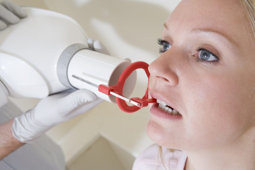
- Preparation: The dental professional will explain the procedure and guide the patient on how to position themselves in the dental chair. Patients are usually seated upright or slightly reclined.
- Placement of the X-ray film or sensor: A small, flat piece of film or a digital sensor is placed in the patient’s mouth, behind the specific tooth or area to be examined. The patient will be asked to bite down gently to hold the film or sensor in place.
- Taking the X-ray: The X-ray machine is positioned adjacent to the patient’s head, targeting the area of interest. The actual exposure time for the X-ray is very brief, usually just a few seconds. During this time, the patient needs to remain still to ensure a clear image is captured.
- Repeat as needed: Depending on the number of angles or areas to be examined, the process may be repeated several times to obtain different views of the teeth and jawbone.
Safety Measures
While Periapical X-rays involve exposure to a small amount of radiation, modern dental practices use techniques that minimize this exposure, such as lead aprons and thyroid collars, which protect the rest of the body. Digital X-ray systems further reduce radiation exposure compared to traditional film X-rays.
Post-X-ray Discussion
After the X-rays are taken, the dentist will review the images and discuss the findings with the patient. This is an excellent opportunity for patients to ask questions and understand the condition of their oral health, particularly in relation to the planned dental implant. The X-ray results play a crucial role in determining the next steps in the implant process, such as the need for bone grafting, the selection of the implant size and type, and the scheduling of the implant surgery.
In summary, Periapical X-rays are a routine and essential part of preparing for dental implants, providing critical information that ensures the best outcomes for the procedure. Patients can expect a quick, straightforward process with minimal discomfort and should take the opportunity to engage with their dental professional about the findings and their implications for the implant plan.
Conclusion
In conclusion, Periapical X-rays stand as an indispensable tool in the realm of dental implant planning, offering a detailed glimpse beneath the surface to ensure that each implant procedure is conducted with the highest degree of precision and care. Their ability to provide comprehensive images of the tooth, root, and surrounding bone structure is paramount in identifying potential issues, assessing bone health, and planning the surgical approach. While they come with certain limitations, these can be effectively managed through skilled interpretation and integration with other imaging modalities. For patients, understanding the process and significance of these X-rays can demystify the initial steps toward dental implant surgery, setting the stage for successful outcomes. By leveraging the detailed insights afforded by Periapical X-rays, dental professionals and patients alike can navigate the implant journey with confidence and clarity.