In the world of modern dentistry, dental implants have emerged as a game-changer, offering durable and natural-looking solutions to tooth loss. However, the key to their success lies in meticulous planning and precision, primarily driven by diagnostic imaging. These images provide vital insights into a patient’s unique oral anatomy, guiding dental professionals in the placement of implants and ensuring long-term success. In this blog post, we will explore the critical role that diagnostic imaging plays in dental implantology, discussing the various techniques used and how they contribute to confident smiles and improved oral health.
The Role of Diagnostic Imaging
Diagnostic imaging plays a crucial role in implantology in several ways:
Assessment of Oral Anatomy:
Diagnostic imaging techniques like X-rays and cone-beam computed tomography (CBCT) provide detailed images of the patient’s oral anatomy. These images reveal the structure of the jawbone, the density of the bone, the location of vital structures like nerves and blood vessels, and the condition of the surrounding teeth and gums. This information is essential for understanding the patient’s unique dental landscape.
Precise Planning:

With a clear view of the patient’s oral anatomy, dental professionals can plan the implant procedure with precision. They can choose the appropriate implant size, shape, and length based on the available bone and the desired outcome. The precise positioning of the implant is critical for both functionality and aesthetics.
Minimizing Complications:
Diagnostic imaging helps identify any potential issues or challenges before the surgery. This allows the dental team to anticipate and address them during the planning phase, reducing the risk of complications during the implant procedure. It helps prevent damage to adjacent teeth, nerves, and blood vessels.
Optimizing Implant Placement:
The images guide the dental surgeon in determining the optimal location and angle for implant placement. This ensures that the implant integrates seamlessly with the natural dental structure, providing stability and support for the prosthetic tooth or teeth.
Long-Term Success:
Diagnostic imaging is instrumental in achieving long-term implant success. It helps in the assessment of bone quality and quantity, ensuring that there is adequate support for the implant. It also aids in monitoring the healing process after the surgery and assessing the stability of the implant over time.
Patient Communication:
Diagnostic images provide a visual representation that can help patients better understand the treatment plan. Dentists can use these images to educate patients about the procedure, expected outcomes, and potential challenges, fostering informed decision-making and peace of mind.
In summary, diagnostic imaging is an indispensable tool in implantology, as it enables dental professionals to assess the patient’s oral anatomy, plan the procedure accurately, minimize complications, and ensure the long-term success of dental implant treatments. It enhances patient care by providing a comprehensive and detailed view of the factors that impact the success of the implant procedure.
Types of Diagnostic Imaging Techniques
Dental implant dentists use various diagnostic imaging techniques to assess a patient’s oral anatomy and plan for successful implant placement. These techniques include:
Dental X-Rays
Dental X-rays are fundamental diagnostic tools that play a pivotal role in the success of dental implants. These two-dimensional images provide valuable insights into a patient’s oral anatomy, bone density, and the proximity of adjacent structures, all of which are critical factors in implant planning. By assessing the condition of the jawbone and identifying potential challenges or complications, dental professionals can make informed decisions regarding implant size, position, and angulation. This precision ensures that implants integrate seamlessly with the natural dental structure, enhancing stability and long-term success. Dental X-rays also aid in minimizing the risk of complications during the implant procedure, such as damage to nerves or neighboring teeth, contributing to a more predictable and satisfactory outcome for patients seeking dental implant solutions.
Cone-Beam Computed Tomography (CBCT)
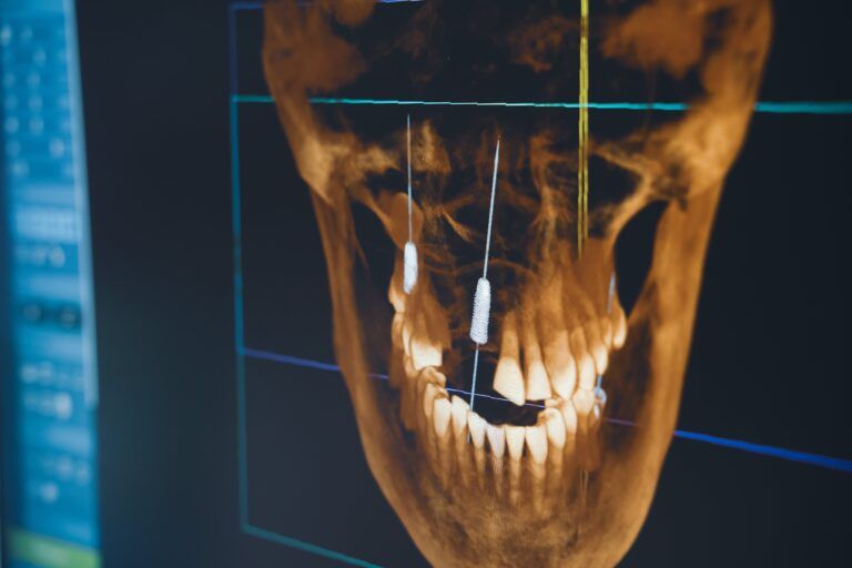
Cone-Beam Computed Tomography (CBCT) scans are a revolutionary advancement in dental imaging that significantly enhances the success of dental implants. Unlike traditional X-rays, CBCT provides detailed three-dimensional images of the patient’s oral and maxillofacial region, offering comprehensive information about bone density, volume, and quality. These precise images are invaluable for implant planning, allowing dental dentists to select the optimal implant size, position, and angle with unparalleled accuracy. CBCT also helps identify the location of vital structures like nerves and blood vessels, reducing the risk of complications during surgery. With this advanced imaging technique, dental professionals can navigate the implant procedure with confidence, ensuring that the implant integrates seamlessly into the patient’s oral anatomy, resulting in a higher likelihood of long-term implant success and patient satisfaction.
Face Hunter

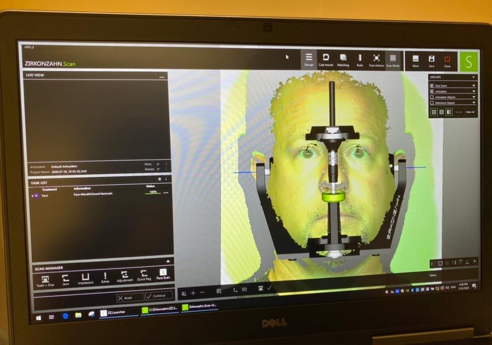
The Face Hunter is a highly advanced tool designed to enhance the success of dental implant procedures. It utilizes state-of-the-art technology to create photo-realistic 3D digitalizations of faces, providing a vital working base for crafting individualized dental prostheses. This technology significantly boosts planning reliability for dental technicians, dentists, and patients by enabling the construction of tooth restorations tailored to each patient’s unique physiognomy. Its intuitive one-click digitalization process and rapid scanning speed of less than 0.3 seconds per face streamline the workflow, making it highly efficient.
Digital Impressions
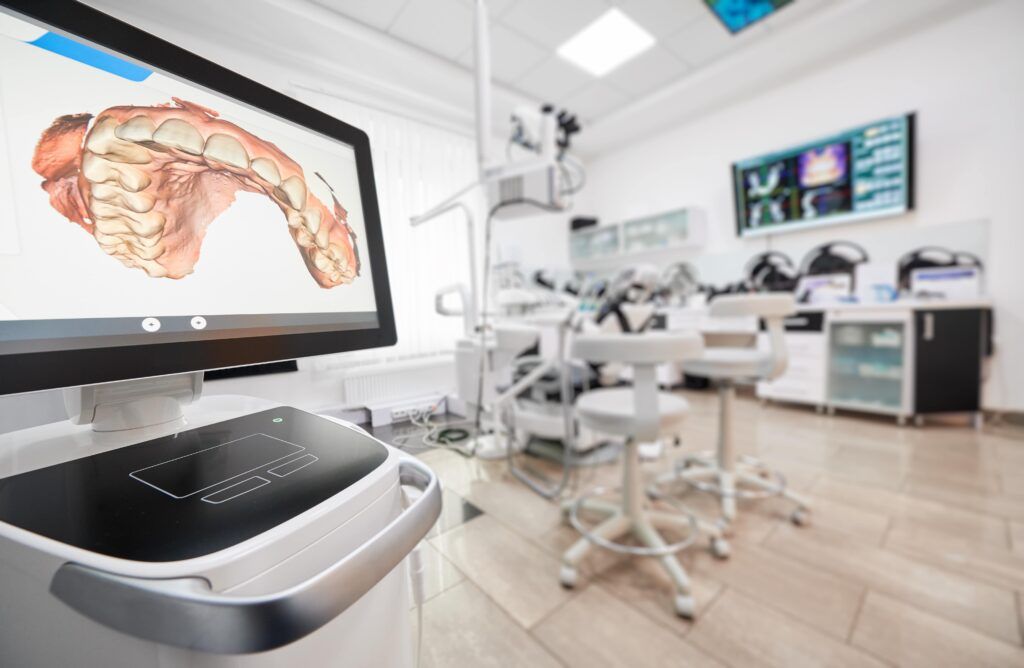
Digital impressions are instrumental in enhancing the success of dental implants. These advanced technologies, such as intraoral scanners, allow dental professionals to create precise three-dimensional digital models of the patient’s oral cavity. While their primary use is for designing prosthetic components like crowns and bridges, these digital impressions also benefit dental implant procedures. They enable accurate measurements and assessments of the implant site, ensuring an optimal fit and alignment of the implant. This precision is essential for achieving both functional and aesthetic results, as it minimizes gaps between the implant and the prosthetic tooth, leading to improved stability and long-term success. Digital impressions also streamline the workflow, reduce the need for traditional, often uncomfortable impression materials, and provide a faster and more efficient implant placement process, ultimately contributing to higher patient satisfaction and dental implant success.
Photographic Documentation
Photographic documentation plays an essential role in ensuring the success of dental implants. By capturing high-quality intraoral and extraoral photographs, dental professionals create a visual record of the patient’s oral condition. These images serve as valuable references throughout the implant treatment process. They help in precise treatment planning, allowing dentists to evaluate the initial condition of the implant site, identify any anomalies, and make informed decisions regarding implant size, positioning, and prosthetic design. Moreover, photographic documentation aids in patient communication, helping individuals better understand their treatment options and expected outcomes. As the implant procedure progresses, these images provide a means of tracking healing, tissue changes, and implant stability, contributing to a well-documented and successful dental implant journey.
How Dental Implant Dentists Utilize Diagnostic Imaging
Dental implant dentists use various diagnostic imaging techniques to plan and execute successful dental implant procedures. Here’s how they typically use these imaging tools:
Assessment of Oral Anatomy:
Dentists begin by using diagnostic imaging, such as X-rays, panoramic radiographs, or CBCT scans, to assess the patient’s oral anatomy. These images provide a detailed view of the jawbone, surrounding structures, and any existing dental issues. They use this information to determine the suitability of the implant site and identify potential challenges or complications.
Treatment Planning:

Based on the diagnostic images, dental implant dentists plan the implant procedure meticulously. They consider factors like bone density, volume, and quality to select the appropriate implant size, type, and placement location. The goal is to ensure optimal stability, support, and aesthetics for the final restoration.
3D Visualization:
CBCT scans offer three-dimensional images that provide an in-depth, 360-degree view of the implant site. Dentists can rotate and manipulate these images digitally, allowing for precise evaluation and planning. This 3D visualization is particularly valuable for complex cases and helps in determining the ideal implant angulation and depth.
Location of Vital Structures:
Diagnostic imaging helps dentists pinpoint the location of vital structures like nerves and blood vessels. This information is crucial for avoiding damage during the implant procedure. By accurately mapping these structures, dentists can adjust the implant placement to minimize the risk of complications.
Prosthetic Planning:
Intraoral scanners and digital impressions create digital models of the patient’s oral cavity, aiding in prosthetic planning. Dentists can design customized crowns, bridges, or other prosthetic components that fit precisely with the implant, ensuring optimal function and aesthetics.
Real-Time Guidance:
Some dentists use guided implant surgery techniques, where they rely on computer software to plan and execute the implant placement with extreme precision. These systems use diagnostic imaging to guide the implant placement in real-time during surgery, minimizing human error.
Monitoring and Follow-Up:
After the implant surgery, diagnostic imaging continues to play a role in the follow-up process. Dentists use X-rays or CBCT scans to assess the healing progress, confirm osseointegration (the fusion of implant and bone), and monitor the long-term stability and health of the implant.
In essence, dental implant dentists use diagnostic imaging as a fundamental tool throughout the entire implant treatment process. It enables them to plan meticulously, minimize risks, optimize aesthetics and functionality, and ensure the long-term success of dental implant procedures for their patients.
Conclusion
In the world of modern dentistry, diagnostic imaging stands as an indispensable pillar of dental implant success. From the initial assessment of oral anatomy to the precise planning of implant placement and prosthetic design, these imaging techniques are the cornerstones of achieving optimal results. They minimize complications, ensure the long-term stability of implants, and foster patient satisfaction. In the realm of dental implantology, diagnostic imaging isn’t just a tool; it’s the key to unlocking confident smiles, improved oral health, and a brighter future for countless individuals seeking the life-changing benefits of dental implants. It exemplifies the commitment of dental professionals to the art and science of their craft, making dental implant procedures safer, more predictable, and ultimately more successful.

